Phi Optics, Inc.  

Champaign,  IL 
United States
https://www.phioptics.com
  • Booth: 634

Phi Optics upgrades existing optical microscopes to provide 4D label-free quantitative imaging of live specimens. Researchers can see live cells and tissues with higher contrast than traditional microscopy modalities. Output data is repeatable and sample invariant, and is suitable for machine learning and automatic screening


 Products

  • CellVista SLIM
    Phi Optics CellVista SLIM upgrades new and existing optical microscopes for 4D quantitative, label-free imaging assays of live cell cultures. Proliferation, viability, cytotoxicity, and more with sub-cellular resolution and no sample preparation...

  • Patented Spatial Light Interference Microscopy (SLIM) is implemented as an add-on to the camera port of your research microscope (Zeiss, Nikon, Leica, Evident) for any magnification and immersion (10X-100X, dry or water/oil and more). SLIM collects live, label-free and quantitative information (morphology, dry mass, RI) with subcellular resolution from milliseconds to days. Phi Optics CellVista SLIM modules provide nanometer and femtogram sensitivity to morphology and mass, high SNR with no speckles background, optical sectioning for 3D tomography. Drug discovery, protein particles, proliferation, viability, cytotoxicity, immune cell killing, and more population assays can be done with sub-cellular resolution. Use regular sample holders (single or multi-wells) or microfluidics with minimal or no sample preparation. Correlative imaging (SLIM and fluorescence) can be acquired with same large field of view, high QE camera for pixel level multi-channel registration to input into latest machine learning models.

  • CellVista GLIM
    Phi Optics CellVista GLIM upgrades new and existing optical microscopes for 4D quantitative, label-free imaging assays of live 3D specimens: organoids, embryos, model animals, plant tissue and more. Sub-cellular resolution with no sample preparation....

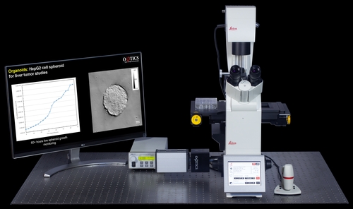
  • Patented Gradient Light Interference Microscopy (GLIM) is implemented as an add-on to the camera port of your research microscope (Zeiss, Nikon, Leica, Evident) for any magnification and immersion (10X-100X, dry or water/oil and more). GLIM collects live, label-free and quantitative information (morphology, dry mass, RI) with subcellular resolution from milliseconds to days. Phi Optics CellVista GLIM modules provide high SNR with no speckles background, optical sectioning for 3D tomography in optically thick specimens: organoids, embryos, small model animals, plant tissue and more. Drug discovery, proliferation, viability, cytotoxicity, immune cell killing, and more assays can be done with sub-cellular resolution. Use regular sample holders (single or multi-wells) or microfluidics with minimal or no sample preparation. Correlative imaging (GLIM and fluorescence) can be acquired with same large field of view, high QE camera for pixel level multi-channel registration to input into latest machine learning models.

  • CellVista AI
    Phi Optics CellVista AI is a live digital staining software option for our SLIM and GLIM modules. CellVista AI will enable 2D and 3D assays with the accuracy and specificity of regular fluorescence, but without phototoxicity and photobleaching...

  • Patented Phase Imaging with Computational Specificity (PICS) uses a neural network to learn from colocalized label-free (CellVista SLIM or CellVista GLIM) and ground-truth fluorescence images and predict where specific fluorophores would bind in an unlabeled specimen – thus providing digital staining (label-free) of the QPI quantitative image. The digital staining masks are used to segment QPI (SLIM or GLIM) images to extract dry mass, morphology and refractive index values for 2D and 3D quantitative assays. Even more, the neural network can extract cell cycle-, cell viability-dependent (and more) features from the nanoscale structure measured in QPI images. 2D and 3D assays can be performed with the accuracy and specificity of regular fluorescence, but without the inconveniences associated with phototoxicity and photobleaching.